"I Thought It Was Just Muscle Pain"... The Disease That Decides Life or Death Within 24 Hours [Kok! Health]
Fatal Outcomes Possible if Misdiagnosed as Myocardial Infarction and Given Antithrombotic Agents
Blood Pressure Management and Aerobic Exercise Recommended After Surgery
The aorta is the largest blood vessel in the body, responsible for carrying blood from the heart to the entire body. It is composed of three layers: the intima, media, and adventitia. A condition called "aortic dissection" occurs when the intima tears, allowing blood to flow between the layers of the vessel wall. Once it occurs, it can rapidly progress to blood flow obstruction, organ ischemia, cardiac tamponade, or aortic rupture, making prompt diagnosis and treatment critically important.

Professor Park Yukyung, a cardiovascular and thoracic surgeon at Soonchunhyang University Bucheon Hospital, stated, "Aortic dissection is a race against time," adding, "If you experience sudden, tearing chest pain accompanied by neurological symptoms such as fainting, paralysis, or altered consciousness, you should not dismiss it as simple muscle pain and must visit the emergency room immediately."
The most characteristic symptom of aortic dissection is sudden, severe chest pain. The pain often starts in the chest and can radiate to the back, abdomen, or lower back. When neurological symptoms are present, it can be difficult to complain of typical chest pain. In such cases, uncontrolled hypertension, loss of pulse in certain areas, and differences in blood pressure between the arms or legs may indicate aortic dissection.
Findings such as widening of the mediastinum between the lungs or an enlarged cardiac silhouette on a chest X-ray can also aid in diagnosis. However, a definitive diagnosis is made through a CT scan, which is used to identify the location of the intimal tear and the extent of the dissection to plan treatment. Echocardiography can be used to check for concomitant aortic valve regurgitation, left ventricular systolic function, and the presence of cardiac tamponade.
Aortic dissections are classified into Stanford type A and type B based on the location of the dissection. Type A involves the ascending aorta and is a life-threatening, ultra-emergency condition that requires immediate surgery upon diagnosis. The ascending aorta is located near the major vessels leading to the heart and brain, so if the dissection progresses, it can result in cardiac tamponade, aortic valve dysfunction, or impaired cerebral blood flow. If surgery is not performed within 24 hours, the mortality rate reaches approximately 25 to 50 percent, indicating a poor prognosis.
Stanford type B, where the dissection is limited to the descending aorta, is generally managed with medical therapy such as blood pressure and pain control as the first line of treatment. However, if there is organ ischemia, persistent pain despite blood pressure control, or a high risk of aortic enlargement or rupture, stent graft placement may be considered. Recently, CT imaging is used to comprehensively evaluate the size of the aorta, the ratio of true lumen to false lumen, the rate of aortic enlargement, and the location of the intimal tear to determine the need for stent treatment. However, stent procedures also carry risks of complications such as retrograde extension of the dissection or paralysis due to spinal cord ischemia, so careful patient selection is essential.
Aortic dissection presents with symptoms similar to acute myocardial infarction, making early differentiation crucial. Both conditions cause chest pain, but myocardial infarction typically involves gradually worsening, tightening or crushing chest pain that may radiate to the left arm or jaw. In contrast, aortic dissection is characterized by sudden, tearing pain that may migrate to the back or abdomen. If aortic dissection is mistaken for myocardial infarction and antithrombotic agents are administered, it can lead to fatal outcomes such as postoperative bleeding, so caution is warranted.
Surgery for type A aortic dissection involves replacing the torn segment of the aorta with a synthetic graft. During the operation, it is necessary to temporarily control blood flow, so protecting the brain and major organs is critical. Techniques such as hypothermia, which reduces the oxygen demand of organs by lowering body temperature, and selective cerebral perfusion, which supplies blood directly to the brain, are utilized. The development of these surgical methods has been reducing the risk of neurological complications.
Depending on the extent of aortic involvement, spinal cord ischemia can cause paralysis of the lower limbs, so preventive measures are also important. If necessary, cerebrospinal fluid drainage can be performed to lower spinal cord pressure, and maintaining blood pressure above a certain level before and after surgery is essential to protect spinal cord blood flow.
Hot Picks Today
 "End of Samsung and SK Hynix’s Dominance?" CXMT Soars 466% on IPO Debut... Founder’s Wealth Jumps 300%
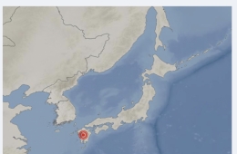
- "Strong Earthquake Causes Partial Collapse of Kyushu Shopping Mall... Many Reportedly Trapped Inside"
- "Voter Numbers Misentered"... Vote Statistics Manipulated in Gimpo, Uiwang, and Jincheon During June 3 Local Elections
- Korea Housing Finance Corporation to Write Off 35% of Principal for Jeonse Fraud Victims Who Repay Remaining Debt in Lump Sum
- Prosecution Requests Arrest Warrant for Entertainment Agency CEO Chagawon on Multi-Billion Won Fraud Charges
Even after surgery or stent procedures, the remaining aorta can dilate or develop further dissection, so ongoing follow-up is necessary. Professor Park emphasized, "Blood pressure management is the most important factor after surgery, and abstaining from smoking, drinking, and avoiding stress is also essential." She added, "Rather than resistance exercises that can cause a sudden spike in blood pressure, aerobic activities such as walking or stationary cycling are helpful for recovery. It is safest to monitor blood pressure changes during exercise as part of a cardiac rehabilitation program and adjust intensity accordingly."
© The Asia Business Daily. All rights reserved. Unauthorized AI training and use prohibited.